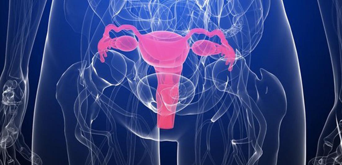
A common type of noncancerous tumor formation known as ‘uterine fibroids’ can be identified during your normal visit to your gynecologist. A pelvic examination will allow your doctor to identify it by feeling the shape and size of the uterus. If your uterus has an irregular shape or is enlarged, then it might be a sign of the presence of a fibroid. Or, the symptoms that you are experiencing can be explained to your doctor for him to know more about it. If, after the examination, your doctor suggests that this looks like a fibroid then certain tests can be performed to confirm the diagnosis. Here’s how uterine fibroids are diagnosed
Ultrasound
Ultrasound is usually the first test that is performed. Most women are already aware of this test from pregnancy. It is not only a reliable procedure but is also safe because no radiation is used in this procedure. Ultrasound makes use of sound waves for creating an image of your ovaries and uterus in around 30 to 60 minutes. Initially, a transducer is used on the abdomen and a conducting gel is used on the skin that makes the skin feel cool and wet. Moving the transducer allows the technologist to take pictures and images.
Then an internal examination is performed where a special ultrasound probe is placed in the vagina. It is inserted like a tampon and is not painful. This takes close-up pictures of the endometrium, uterus, and ovaries.
Lab tests
In case of abnormal and heavy bleeding, one’s doctor may suggest them some lab tests for investigating the cause. To diagnose the potential causes, you may have to take a complete blood count for determining the exact cause of anemia. Other blood tests can be performed for ruling out thyroid and bleeding disorders.
Saline hysterosonography
Saline hysterosonography is also an ultrasound procedure that does not use any radiation. This procedure helps to better visualize the internal examination. This method is great at identifying submucosal fibroids and polyps.
This exam is usually performed after the woman finished her menstrual period and takes about half an hour. Saline hysterosonography uses a small catheter that is inserted via the cervix, where a small balloon is then inflated for holding it in place. The uterus is then injected with sterile saline and ultrasound images are taken. Cramps similar to menstrual cramping may be experienced during the procedure and even after the procedure has been performed and this is normal.
MRI
Magnetic Resonance Imaging (MRI) despite being expensive, is capable of forming a very detailed image that shows the size, number, and exact position of the fibroid. Not all women will require an MRI. This procedure also doesn’t use radiation instead uses a special and large magnet for taking the pictures.
The total time taken to perform this process is around 45 to 60 minutes, the patient is asked to remain still during this time. An intravenous line is placed in the arm before starting with the procedure, it is injected with contrast material for taking pictures of the pelvic area. Then the patient is laid on a moveable bed and is passed through a big magnet, shaped like a donut.
A radiologist then reviews the pictures and reports to the doctor about what he found.
Hysteroscopy
It is another method to look inside the uterus. This procedure can be performed in an operating room or even in a doctor’s office. The test is completed in 30 minutes.
During the test, a speculum is placed in the vagina while the patient lies on their back and their feet are in gynecology stirrups. A telescope that is very long and is known as a hysteroscope is then inserted into the uterine cavity through the cervix. CO2 and sterile saline can be injected through the hysteroscope to inflate the cavity and create an ideal image. The formed images are displayed on the TV monitor. Mild cramps can occur during the hysteroscopy. The discomfort can be avoided by taking ibuprofen an hour before going into the procedure.


