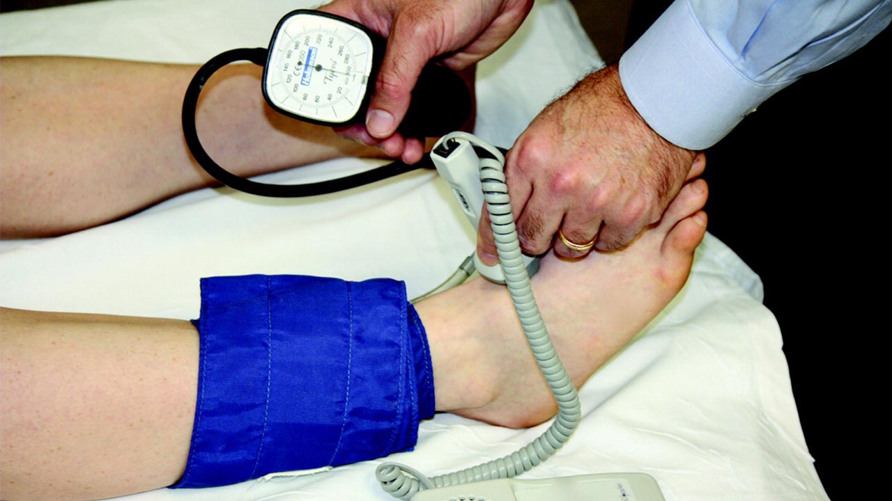
If we are talking about noninvasive tests, a Doppler ultrasound is worth mentioning. This procedure measures the blood flowing through the blood vessels by using high-frequency sound waves. These sound waves bounce off the red blood cells that are circulating in the blood for creating images.
Doppler screening is known for the diagnosis of many conditions that include clotting of blood, valves that function poorly in the leg veins hence causing the blood to accumulate in the legs, blockage of arteries, and narrowing of the arteries in your body. Peripheral arterial disease tops the list of diseases that can be diagnosed by Doppler screening.
Types of Doppler Ultrasounds
Doppler ultrasound tests are of different kinds; these can be listed as:
Color Doppler: Such a Doppler test uses a computer to convert sound waves into different colors. These colors then indicate the direction and speed of the flow of blood.
Power Doppler: It is an enhanced type of color Doppler. It provides greater detail of the flowing blood as compared to the standard color Doppler. However, it is not capable of showing the direction to where the blood flows, which is very important to know in some cases.
Spectral Doppler: This type of Doppler test is great at showing information regarding the blood flow on a graph instead of some color pictures. This method is capable of showing the blockage of blood vessels.
Duplex Doppler: This type of Doppler test will use standard ultrasound, for creating images of the vessels, and a computer to turn those images into graphs just as the spectral Doppler.
Continuous wave Doppler: This test is the most accurate one for measuring the speed of blood flow. It continuously sends and receives sound waves for its work.
Functioning of the Doppler ultrasound
It can also be called an imaging test. The difference between a regular ultrasound and a Doppler ultrasound is that the Doppler ultrasound is capable of showing the blood moving through the vessels but the regular ultrasound can only produce images and does not show the blood flowing through vessels.
Doppler ultrasound measures the sound waves that reflect from objects that are in motion, i.e. red blood cells. Hence, makes use of the Doppler Effect.
Usually includes the following steps:
⦁ The patient is laid on the table and exposed to the area that is being screened
⦁ A special gel is then spread over the exposed area
⦁ Transducer is moved over that area
⦁ This wand-like device will then send sound waves into the patient’s body
⦁ A change in the pitch of sound waves occurs due to the moving blood cells, a pulse-like or swishing sound may also be heard during this procedure
⦁ These waves are then recorded and the recorded data is then converted into images and graphs on a computer
⦁ After the test has been completed, the gel will be wiped off the body
⦁ The total time taken for the test to complete will be 30-20 minutes
Preparation for the screening
Several things need to be followed to prepare for the Doppler screening, these include the removal of clothes and any type of jewelry that is present on or around the area that is going to be tested, the patient should avoid taking products that consist of nicotine for almost 2 hours before the test happens or else the blood vessels will be narrowed because of nicotine and in certain types of Doppler testing, one will be asked to fast for a few hours before the test happens.
Risks involved?
Doppler screening is considered safe even during pregnancy. There is no known risk associated with the Doppler Screening. If the results of the Doppler screening are not normal, it would potentially mean that you might have a blocked artery or a clot in any artery, your blood vessels have narrowed, your blood flow is abnormal or there is a balloon-like bulge known as aneurysm in your arteries. Aneurysm causes the vessels to become thin and stretched. The thinning of the walls can cause the vessels to rupture, which can be life-threatening.


