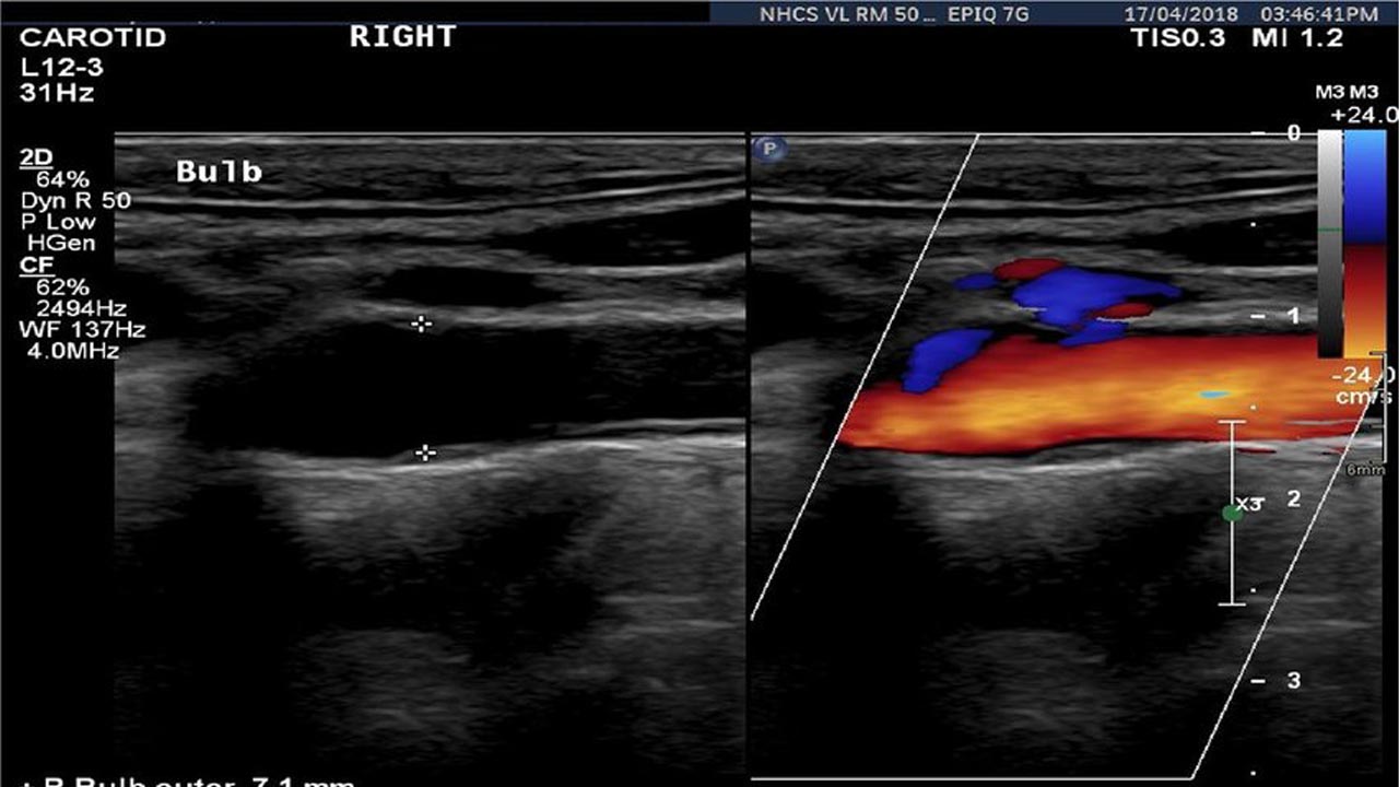
Duplex Ultrasound is a non-invasive evaluation of your blood flow and involves high-frequency sound waves. These high-frequency sound waves help look at the structure of leg veins and the speed at which the blood flows within them. The word ‘duplex’ stands for the two modes of ultrasound that are used, i.e., B-mode and Doppler. While performing the duplex ultrasound, two displays are available. The B-mode will help to get an image of the vessel that is intended to be studied. In comparison, the Doppler evaluates the direction and the velocity of the blood flowing in that vessel.
Duplex Ultrasound is an integral part of medical diagnosis and therapy. It helps vascular surgeons with the diagnosis of the problem and also about how to make a treatment plan. In such conditions, accuracy is very crucial. Ultrasound tests should be conducted in officially recognized and authorized vascular laboratories to ensure the results’ accuracy and liability.
Advantages of Duplex Ultrasound
- Comprehensive
- Mobile
- Well tolerated
- Non-invasive
- No exposure to nephrotoxic contrast or radiation
- Highly operator dependent
Procedure
The arteries and veins in the carotid, leg, and arm can be studied by ultrasound. Doctors tend to examine the arteries, veins, and abdominal aorta via ultrasound. Clots or refluxes can also be detected using this method.
These types of examinations are very easy for patients as they are painless. They are, however, recommended in a certified vascular laboratory. A bed with a few pieces of equipment is all needed to perform an ultrasound. Patients can wear whatever clothing they are comfortable with. The facility provides disposable shorts and gowns. The entire procedure takes up to 30 minutes. Very little preparation is required for duplex ultrasound, and patients may be required to fast if they have an abdominal examination.
Necessary Equipment
- Ultrasound machine with duplex capabilities
- Probe with piezoelectric crystals
- Transducers
The transducers used should be enabled for varying focus and direction and should be capable of adjustment with the resolution and depth of ultrasound waves. The equipment used should be compatible with multiple transducers for varying advantages as follows:
- Linear array transducer: Used for anatomic mapping of the arteries and veins
- Low-frequency transducer: Beneficial for the visceral vessels and abdominal imaging
- Harmonic imaging transducer: Particularly used for high depths and providing increased resolution.
Duplex ultrasound typically has no side effects or complications associated with its examination procedures. But, some procedures may pose a degree of risk.
Significance of Duplex Ultrasound
The procedure, duplex ultrasound, is a very significant clinical tool. The continuous expansion in the application of this procedure is due to its affordability, safety conditions, probability, accuracy, and tolerance. It is now readily available in many healthcare facilities. Hence, it is very important for nurses, healthcare workers, surgeons, and internees to realize its value and should utilize it as a technique that is a very portable and effective method.